|
Physics of biology and medicine
Reference:
Pashovkin, T.N., Sadikova, D.G. (2024). Effect of continuous and modulated ultrasound on fish neurons. Physics of biology and medicine, 1, 41–54. . https://doi.org/10.7256/2730-0560.2024.1.71004
Effect of continuous and modulated ultrasound on fish neurons.
Pashovkin Timofei Nikolaevich
ORCID: 0000-0001-9697-9230
Doctor of Biology
Leading Researcher; Institute of Cell Biophysics of the Russian Academy of Sciences - a separate division of the FITC PNCBI RAS
29 Nauki ave., 48 sq., Pushchino, Moscow region, 142290, Russia

|
pashovkin@mail.ru
|
|
 |
Other publications by this author
|
|
|
Sadikova Diana Gablel'fartovna
PhD in Physics and Mathematics
Researcher; Institute of Cell Biophysics of the Russian Academy of Sciences - a separate enterprise of the FITC PNCBI RAS
142290, Russia, Pushchino, Studentskiy lane, 16, sq. 54
|
|
sdg7@list.ru
|
|
|
|
DOI: 10.7256/2730-0560.2024.1.71004
EDN: QYEYTI
Received:
11-06-2024
Published:
30-08-2024
Abstract:
Currently, transcranial ultrasound stimulation (TUS) is being intensively developed as a new non-invasive method of neuromodulation. A convenient model for demonstrating ultrasonic neuromodulation is the nervous system of fish. Experiments have been carried out on Goldfishes. We have recorded general swimming reaction and turning reaction of fishes in the special chamber which bottom had been divided into sectors. We observed decrease of general swimming reaction and turning reaction after influence of continuous ultrasonic waves of therapeutic intensities (f = 0.88 MHz), when intensity was more than 0.7 W/cm2, and increase of these responses at intensities less than 0.1 W/cm2. Application of modulated ultrasonic fields as an acting factor produced changes of activity of fishes dependent on a modulating frequency. The action spectra have been received using an amplitude modulation (AM) of low frequency. This spectra reflect the work of the whole brain (tests of change of a general swimming reaction of fishes), and the work of identified Mauthner’s neuron, that is responsible for turning response of fishes. The action spectrum for Mauthner’s neuron is more expressed and contains three kinds of frequencies by the action on fishes activity: frequency of activation (8 Hz), partially depressing (6, 10 Hz) and neutral (3, 7, 9 Hz). Spectra are received at equienergy action АМ of ultrasonic sound irrespective of a modulating frequency (porosity = 2) and spatial average and temporal average intensities of 0.35 W/cm2. From an action spectrum we can conclude, that on one modulating frequency effects of a carrier frequency are relaxed, and on others strengthen. This approach can find application in ultrasonic therapy when it is necessary to make ultrasonic action more effective and to decrease potential hazard of action due to the cavity action.
Keywords:
ultrasound, modulation, action spectrum, Mauthner neuron, neuromodulation, frequency modulation, motor activity, turning response, activation, suppression
This article is automatically translated.
You can find original text of the article here.
Introduction. Any effects that lead to changes in the electrical activity of neurons are central to basic research and are important for the clinical treatment of neurological disorders. In model animals and humans, visualization of changes in the activity of both individual neurons and neural activity on the scale of the whole brain is the main way to develop neurobiological technologies. The available electrical and optical methods, as a rule, do not work at this scale due to their inherent physical limitations. An alternative to these methods is the use of ultrasound, which interacts with brain tissues with a fundamental resolution of about 100 microns and a time resolution of 1 ms. Transcranial ultrasound stimulation (TUS) is currently being intensively developed as a new, non-invasive neuromodulation method [3]. TUS has a higher spatial resolution than transcranial magnetic stimulation (TMS) [1] or direct electric current stimulation [2], which are currently used in practice. Almost a hundred years ago, it was first recognized that ultrasound modulates the electrical activity of cells. Since then, ultrasound neuromodulation in the brain, in the peripheral nervous system of humans and model organisms has been widely covered in the literature [3-10]. Despite the long period of research. The fundamental cellular, molecular, and mechanical foundations of ultrasonic neuromodulation still remain largely unknown [3-11]. It is noted that ultrasound creates excitatory [12,13] and/or inhibitory effects [14.15] depending on the system under study and the parameters of the stimulus [3, 13, 14]. Thus, stimulation of the motor cortex of the brain in animals can induce electromyographic (EMG) signals in the corresponding muscles of the forelimb, hindlimb or tail, which indicates the possibility of TUS to induce action potentials [15.16]. The inhibitory effects of TUS are manifested in a long-term weakening of synaptic transmission [17]. It is believed that these opposite effects on the activity of neurons are due to differences in the place of stimulation, intensity, frequency of ultrasound and other parameters [18, 28]. Ultrasound can have combined thermal and mechanical effects on neurons [19, 20]. Thermal and cavitation effects of ultrasound, although effectively used to destroy tissue or temporarily open the blood-brain barrier [21], require the use of greater power, frequency and/or duration of exposure than those commonly used for neuromodulation [3]. Due to compression and expansion in the ultrasonic wave, non-selective currents can occur in cell membranes that change the electrical activity of cells [3.11]. Due to the creation of shear stresses in the cell membrane, which increase the tension of the membrane and the appearance of geometric deformation of the lipid bilayer, ultrasound can activate mechanosensitive ion channels [3, 13, 20-26]. Ultrasound can have both thermal and mechanical effects on the field of research in brain tissues. However, the temperature increase will be insignificant at low ultrasound intensities [27], and the forces of acoustic radiation have a mechanical effect without causing cavitation [13]. From the point of view of biophysics, mechanosensitive channels react to the mechanical action of ultrasound [28]. For example, changes in the conductivity of channels may be associated with changes in the shape of channels. Ultrasound can do this due to the appearance of shear stresses in the membranes, or by changing the membrane tension when the pressure in the ultrasonic wave changes. These changes widen the channel and make it more cylindrical in the plane of the membrane when opened [28]. They are energetically favorable in the presence of membrane tension, and this leads to a tension-dependent energy difference between the states, which contributes to the opening of the channel [29,30]. In laboratory conditions, the use of modulated ultrasonic waves can make it possible to identify neural structures that give a frequency-dependent response to external periodic exposure, to identify frequencies at which changes in functional activity will be maximum. Also, in neurobiological research, the choice of an object is important. In this regard, the Mautner neurons (MN) of bony fish and amphibians serve as a convenient object. They have a significant number of afferent inputs and control the swimming activity of fish by controlling the movement of the caudal fin, initiating a unilateral stroke of the caudal fin, the main mover of fish. This makes it possible to judge functional changes in neurons by the behavior of an animal and to influence it in a targeted manner by various experimental influences. MN are two giant cells in the medulla oblongata of most bony fish and amphibian larvae. They are innervated from the vestibular apparatus through the VIII nerve (Fig.1). In turn, MN contralaterally innervates spinal motor neurons, which control the muscles of the trunk. This point of view has been confirmed by numerous experimental data [31-36]. 
Fig. 1. The layout of Mautner neurons in fish. One of the aspects of ultrasound application is the induction of rapid functional changes with minimal manifestation of nonspecific reactions of the nervous tissue. The presence of characteristic frequencies in the electrical activity of neurons suggests that exposure to pulse-modulated ultrasound with appropriate pulse repetition frequencies can cause frequency-dependent changes in the functional state of this tissue. When using ultrasound, as a rule, a significant number of different nerve endings are located in the area of exposure. In the case of changes in their functioning under exposure, it can be assumed that various biochemical processes in organs and tissues innervated by these endings and, consequently, their functional state may also change to varying degrees. This can be most clearly manifested when exposed to identified, specialized neurons responsible for certain functional states of an organ, tissue, and the body as a whole.
The aim of the work is to study the effect of continuous and pulse modulated ultrasound on the morphofunctional state of brain neurons and identified central neurons of vertebrates – Mautner neurons of goldfish. The priority goal of the work is to obtain action spectra for neurons. But in order to obtain them, it is necessary to know how continuous ultrasound affects neurons, since it is used as a factor with a carrier frequency, which is superimposed with modulation frequencies. Therefore, the final effect is the total effect of continuous ultrasound and modulation frequencies. Materials and methods. The activity of brain neurons and functional activity of MN was assessed by the behavior of fish in an annular chamber [31], counting the number of sectors and turns initiated by activation of MN for 10 minutes. The annular chamber consisted of a circular channel with a width of 20 mm and a water level height of 30 mm. The bottom of the chamber was divided into 8 sectors. In this camera, the fish moves in a circle, periodically changing the direction of movement, making turns. Thus, the speed of its movement and the frequency of turns were quantified. The re-movement of the fish around the ring and the turns of the fish are the standard motor reaction of the fish. The test of motor activity consisted in placing the fish in a camera, and measuring the number of sectors traversed and the frequency of turns or changes of direction of movement. Testing in all experiments was carried out for 10 minutes at the same time, since the motor activity of fish depends on the time of day at which the test is performed. Irradiation of MN goldfish was carried out on a separate installation, the block diagram of which is shown in Fig. 3, using a focusing ultrasonic emitter connected to a therapeutic generator UZT-1.01F with a carrier frequency of 0.88 MHz with an average spatial and temporal intensity in the range 0.1 - 1 W/cm2. The irradiation zone of the fish's head was located in the central part of the focal area, with a length of 1 cm and a focal spot radius of 2.5 mm. The ultrasound intensity was evaluated using a differential thermocouple calibrated by ultrasound intensity. The spatial distribution of intensities in the focal area was determined by the paint/paper method, represented as a 2- and 3-dimensional image (Fig.2). The total irradiation time was varied in the range from 15 seconds to 20 minutes. The G6-28 generator was used as a modulator. The fish was fixed in a special thermostatically controlled chamber at a distance of 7 cm from the surface of the radiator. (Fig. 3) 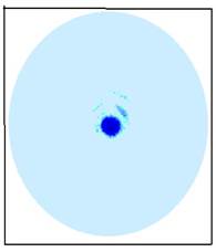 A A 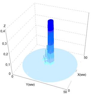 B B
Fig.2. Visualization of ultrasound intensity distributions in the focal area of the emitter. A is a two-dimensional image, B is a three–dimensional image, where Z is the ultrasound intensity scale, W/cm2. 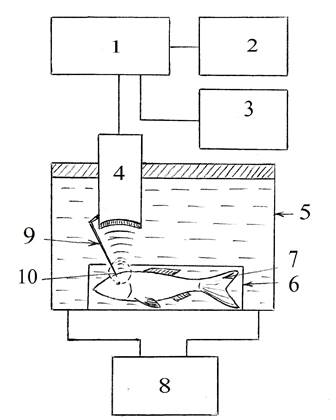
Fig. 3. A block diagram of an installation for ultrasound exposure to fish neurons. 1. Ultrasound generator, 2 – oscilloscope, 3 - time relay, 4 – emitter, 5 - external camera, 6 - internal camera, 7 - object (fish), 8 – thermostat, 9 - focus indicator, 10 - irradiation area. The dubbing was performed at a temperature of 18 ° C. The temperature in the chamber was measured before and immediately after irradiation. The efficiency of the thermostating was no worse than 18+ 0.3 ° C. Morphological changes in MN were studied using a JEM-100B electron microscope after a standard procedure for fixing ultrathin sections of nervous tissue [32]. Research results and discussion. Figure 4 shows changes in the motor activity of fish under the influence of continuous ultrasound with an intensity of 0.1 W/cm2. The figure shows the stimulating effect of low-intensity ultrasound. Figure 5 shows the inhibition of the motor activity of fish under the influence of ultrasound with an intensity of 1.0 W/cm2. This oppression is reversible and disappears: 10 minutes after voicing for 30 seconds, and 30-40 minutes after voicing for 5 minutes. The suppression of activity becomes irreversible after 20 minutes of voicing, with a fatal outcome. 
Fig.4. Stimulation of the fish's rotary reaction (the reaction of the Mautner neuron) depending on the time of exposure to low-intensity ultrasound: I SATA = 0.1 W/cm2, f = 0.88 MHz, t = 18 o C, 100% control level. After suppression by ultrasound with an intensity of 1 W /cm2 and an exposure time of 5 minutes, a rapid restoration of the functional activity of fish is observed, followed by ultrasound exposure with an intensity of 0.1 W /cm2, carried out for 5 minutes. Figure 6 shows the change in the motor activity of fish relative to the magnitude of the effect of continuous ultrasound, under the influence of amplitude-modulated ultrasound in the modulation frequency range 2-14 Hz, with a depth of 2, a modulation depth of 100%, and an irradiation time of 5 minutes.  but but
 b b
Fig. 5. The dependence of fish swimming activity on the time of exposure to continuous ultrasound: a) suppression of swimming activity, b) suppression of the rotary reaction, A) an area of high mortality of fish (more than 90%). I SATA = 1 W/cm2, f = 0.88 MHz, t = 18 oh S It can be seen (see Fig. 6a, b) that the maximum inhibition of motor activity is observed at modulation frequencies of 6 and 10 Hz, and the maximum activation is at a modulation frequency of 8 Hz. Moreover, the rotational reaction of fish changes to a greater extent, while the overall motor reaction changes in the same way, but with with a smaller amplitude. It follows that the identified MN is more susceptible to the action of amplitude-modulated ultrasound than the totality of other neural structures. Functional responses to ultrasound exposure with these modulation frequencies are characteristic both for individual specific neurons and for the neuron system as a whole. The action spectra are given taking into account the effects of the carrier frequency (these effects are subtracted from the spectrum). Moreover, the activation effects caused by a modulation frequency of 8 Hz were canceled by a frequency of 10 Hz, with equal-energy exposure. In this example, one can see the possibility of stabilizing the functional state of the Mautner neuron of fish by using only modulation modes of ultrasonic exposure. Ultrastructural analysis of electron microscopic images showed that, compared with the norm (Fig.7), voicing MN with ultrasound with an intensity of 1 W/cm2 for a time from 10 to 20 minutes leads to destructive changes in the ultrastructure of MN, expressed in the stratification of the myelin sheath and ruptures in the axolemma. This is also accompanied by complete desolation of axosomatic synapses and a general decrease in synaptic vesicles. These morphological changes correlate with the behavior of goldfish in the annular chamber, associated with a decrease in the functional activity of MN after exposure to ultrasound, with an intensity of 1 W/cm2. Ultrasound does not cause visible changes in the myelin sheath or axolemmas of presynaptic fibers when exposed for 5 minutes (ultrasound intensity is average in space and time – ISATA), however, it causes a change in the cytoplasm of MN. These changes consist in the formation of crystal-like structures inside the nucleus (Fig. 8). Similar changes are observed when exposed to pulse-modulated ultrasound with modulation frequencies of 6 and 10 Hz. Crystal-like structures are formed due to the rearrangement of neurofilaments present in the cytoplasm.  But But
 B B
Fig. 6. Dependence of changes in the motor activity of goldfish under the influence of modulated ultrasound on the frequency of modulation: A) rotary reaction, B) general motor activity. I SATA = 0.35 W/cm2 (1/2 intensity of continuous ultrasound = 0.7 W/cm2 – borehole = 2) , f = 0.88 MHz , t = 5 min. 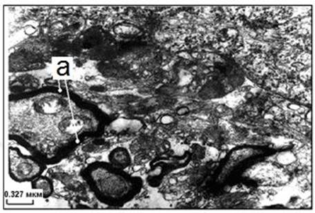
Fig. 7. The structure of goldfish brain tissues before ultrasound exposure. a) the myelin sheath. 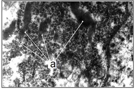
Fig. 8. Formation of neurofilament bundles in the brain tissues of goldfish after ultrasound exposure: a) neurofilament bundles. I SATA =1 W/cm2, f = 0.88 MHz, t = 5 min. An essential fact for therapy is that the sounding of fish with ultrasound of low therapeutic intensity 0.1 W/cm2 leads to the restoration of the structural organization of neurons and the resorption of neurofilament bundles. The effects of continuous ultrasound depend mainly on the mechanisms of biological action (mechanical, thermal, cavitation). With an increase in the intensity of exposure, one of these mechanisms mainly works. As a rule, with increasing intensity, suppression of the functional state of neurons is observed, correlating with morphological changes in the structures of neurons. Mechanosensitive ion channels of various types can be one of the targets of modulated ultrasonic exposure. Thus, their activation can be due to an increase in the tension of the cell membrane or geometric deformation of the lipid bilayer. Such channels may include mechanosensitive ion channels (K2Ps), Piezo1, MEC-4, TRPA1, Msc, and voltage-controlled Na+ and Ca 2+ channels. To date, it is not known exactly how ultrasound affects the activity of these channels. The effects of modulated ultrasound on neurons may differ significantly from the effects of continuous ultrasound. The presence of active frequencies leads to the transformation of biological effects. At frequencies of activation or suppression of functional activity, effects exceeding the amplitude of the effects of continuous ultrasound can be obtained, with the possibility of changing the sign of the effect with a change in intensity. From the data presented in the article, it can be seen that when exposed to modulated ultrasound with equal energy parameters, frequency-dependent effects of stimulation and suppression of neuronal activity can be obtained at significantly lower energies in the ultrasonic beam than when exposed to continuous ultrasound. Thus, it is possible to significantly reduce the intensity of ultrasound exposure in physiotherapy while maintaining the magnitude of biological effects when using modulated ultrasonic fields. The possibility of controlling the functional state of neurons by modulated ultrasound is shown, using various parts of the action spectra obtained for both individual neurons and complex structures of fish neurons for correction.
Conclusions. 1. For fish neurons, the time ranges of the formation of stimulation effects at an ultrasound intensity of less than 0.1 W/cm2 and the effects of inhibition of motor activity of fish at an intensity of 1 W/cm2 are shown. 2. It is shown that it is possible to restore the functional activity of fish using ultrasound stimulation with an intensity of 0.1 W/cm2 immediately after the depressing effect of ultrasound with an intensity of 0.7 - 1.0 W/cm2. 3. For the first time, acoustic action spectra in the modulation frequency range of 2-14 Hz for fish brain neurons, including identified Mautner neurons, are shown. 4. It is shown that it is possible to reduce the risk of ultrasonic negative effects while maintaining the magnitude of the biological effect when using modulated ultrasound. 5. For the first time, the possibility of controlling the sign of a biological effect on fish neurons using equal-energy exposure with modulated ultrasound has been shown.
References
1. De Deng, Z., Lisanby, S. H., & Peterchev, A. V. (2013). Electric field depth-focality tradeoff in transcranial magnetic stimulation: simulation comparison of 50 coil designs. Brain Stimul., 6, 1–13. doi:10.1016/j.brs.2012.02.005
2. Reinhart, R. M. G., Woodman, G. F., & Posner, M. I. (2015). Enhancing long-term memory with stimulation tunes visual attention in one trial. Proc. Natl. Acad. Sci. USA, 112, 625–630. doi:10.1073/pnas.1417259112
3. Blackmore, J., Shrivastava, S., Sallet, J., Butler, C.R., & Cleveland, R.O. (2019). Ultrasound neuromodulation: A review of results, mechanisms and safety. Ultrasound Med. Biol., 45, 1509–1536.
4. Harvey, E.N. (2019). The effect of high frequency sound waves on heart muscle and other irritable tissues. Am. J. Physiol., 91, 284–290.
5. Fry, F.J., Ades, H.W., & Fry, W.J. (1958). Production of reversible changes in the central nervous system by ultrasound. Science, 127, 83–84.
6. Downs, M. E., et al. (2018). Non-invasive peripheral nerve stimulation via focused ultrasound in vivo. Phys. Med. Biol., 63, 035011–11.
7. Munoz, F., Aurup, C., Konofagou, E. E., & Ferrera, V. P. (2018). Modulation of brain function and behavior by focused ultrasound. Curr. Behav. Neurosci. Rep., 5, 153–164.
8. Kamimura, H. A. S., et al. (2016). Focused ultrasound neuromodulation of cortical and subcortical brain structures using 1.9 MHz. Med. Phys., 43, 5730–5735.
9. Tufail, Y., et al. (2010). Transcranial pulsed ultrasound stimulates intact brain circuits. Neuron, 66, 681–694.
10. Fini, M., & Tyler, W. J. (2017). Transcranial focused ultrasound: A new tool for non-invasive neuromodulation. Int. Rev. Psychiatry, 29, 168–177.
11. Naor, O., Krupa, S., & Shoham, S. (2016). Ultrasonic neuromodulation. J. Neural Eng., 13, 031003.
12. Lee, W., Kim, H. C., Jung, Y., Chung, Y. A., Song, I. U., Lee, J. H., et al. (2016). Transcranial focused ultrasound stimulation of human primary visual cortex. Sci. Rep., 6, 1–12. doi:10.1038/srep34026
13. Yoo, S., Mittelstein, D. R., Hurt, R. C., Lacroix, J., & Shapiro, M. G. (2022). Focused ultrasound excites cortical neurons via mechanosensitive calcium accumulation and ion channel amplification. Nat. Commun., 13, 493. doi:10.1038/s41467-022-28040-1
14. Dallapiazza, R. F., Timbie, K. F., Holmberg, S., Gatesman, J., Lopes, M. B., Price, R. J., et al. (2018). Non-invasive neuromodulation and thalamic mapping with low-intensity focused ultrasound. J. Neurosurg., 128, 875–884. doi:10.3171/2016.11.JNS16976
15. Oh, S. J., Lee, J. M., Kim, H. B., Han, S., Bae, J. Y., Hong, G. S., et al. (2019). Ultrasonic neuromodulation via astrocytic TRPA1. Curr. Biol., 29, 3386–3401.e8. doi:10.1016/j.cub.2019.08.021
16. Duque, M., Lee-Kubli, C. A., Tufail, Y., Magaram, U., Patel, J., Chakraborty, A., et al. (2022). Sonogenetic control of mammalian cells using exogenous transient receptor potential A1 channels. Nat. Commun., 13, 600. doi:10.1038/s41467-022-28205-y
17. Niu, X., Yu, K., & He, B. (2022). Transcranial focused ultrasound induces sustained synaptic plasticity in rat hippocampus. Brain Stimul., 15, 352–359. doi:0.1016/j.brs.2022.01.015
18. Dell’Italia, J., Sanguinetti, J. L., Monti, M. M., Bystritsky, A., & Reggente, N. (2022). Current state of potential mechanisms supporting low intensity focused ultrasound for neuromodulation. Front. Hum. Neurosci., 16, 1–23. doi:10.3389/fnhum.2022.872639
19. Dalecki, D. (2004). Mechanical bioeffects of ultrasound. Annu. Rev. Biomed. Eng., 6, 229–248.
20. O’Brien, Jr. W. D. (2007). Ultrasound-biophysics mechanisms. Prog. Biophys. Mol. Biol., 93, 212–255.
21. Choi, J. J., Pernot, M., Small, S. A., & Konofagou, E. E. (2007). Noninvasive, transcranial and localized opening of the blood-brain barrier using focused ultrasound in mice. Ultrasound Med. Biol., 33, 95–104.
22. Tufail, Y., Yoshihiro, A., Pati, S., Li, M. M., & Tyler, W. J. (2011). Ultrasonic neuromodulation by brain stimulation with transcranial ultrasound. Nat. Protoc., 6, 1453–1470.
23. Kubanek, J., Shukla, P., Das, A., Baccus, S. A., & Goodman, M. B. (2018). Ultrasound elicits behavioral responses through mechanical effects on neurons and ion channels in a simple nervous system. J. Neurosci., 38, 3081–3091.
24. Kubanek, J., et al. (2016). Ultrasound modulates ion channel currents. Sci. Rep., 6, 24170.
25. Prieto, M. L., Firouzi, K., Khuri-Yakub, B. T., & Maduke, M. (2018). Activation of Piezo1 but not NaV1.2 channels by ultrasound at 43 MHz. Ultrasound Med. Biol., 44, 1217–1232.
26. Oh, S.-J., et al. (2019). Ultrasonic neuromodulation via astrocytic TRPA1. Curr. Biol., 29, 3386–3401. e8.
27. Tyler, W. J. et al. (2008). Remote excitation of neuronal circuits using low-intensity, low-frequency ultrasound. PLoS One, 3, e3511–e11.
28. Blackmore, D. G., Razansky, D., & Götz, J. (2023). Ultrasound as a versatile tool for short-and long-term improvement and monitoring of brain function. Neuron, 111, 1174–1190. doi:10.1016/j.neuron.2023.02.018
29. Ye, J. et al. (2018). Ultrasonic control of neural activity through activation of the mechanosensitive channel MscL. Nano Lett., 18, 4148–4155.
30. Brohawn, S. G., Campbell, E. B., & MacKinnon, R. (2014). Physical mechanism for gating and mechanosensitivity of the human TRAAK K+ channel. Nature, 516, 126–130.
31. Moshkov, D.A., Podolsky, I.Ya., Kashapova, L.A., Tiras, N.R., Masyuk, L.N., Muzafarova, L.N., & Bolotnova, G.P. (1982). Quantitative characterization of locomotor activity in goldfish as a possible indicator of the state of Mauthner neurons. J. evolution. bioch. and physiol., 2, 155–160.
32. Tiras, N.R., & Moshkov, D.A. (1985). Electron microscopic study of disruption of inhibitory transmission in afferent connections of Mauthner neurons. Cytology, 1, 40–45.
33. Eaton, R.C., Lavender, W.A., & Wieland, C.M. (1982). Alternative neural pathways initiate fast-start responses following lesions of the mauthner neuron in goldfish. J. Comp. Physiol., 145(4), 485–496. doi:10.1007/BF00612814. S2CID 8529312
34. Korn, H, & Faber, D.S. (2005). The Mauthner cell half a century later: a neurobiological model for decision-making?" Neuron, 47(1), 13–28. doi:10.1016/j.neuron.2005.05.019
35. Eaton, R.C., Bombardieri, R.A., & Meyer, D.L. (1977). The Mauthner-initiated startle response in teleost fish. The Journal of Experimental Biology, 66(1), 65–81. doi:10.1242/jeb.66.1.65
36. Zottoli, S.J., & Faber, D.S. (2000). The Mauthner Cell: What Has It Taught Us? Neuroscientist, 6, 26–38. doi:10.1177/107385840000600111
Peer Review
Peer reviewers' evaluations remain confidential and are not disclosed to the public. Only external reviews, authorized for publication by the article's author(s), are made public. Typically, these final reviews are conducted after the manuscript's revision. Adhering to our double-blind review policy, the reviewer's identity is kept confidential.
The list of publisher reviewers can be found here.
The article "The effect of continuous and modulated ultrasound on fish neurons", sent to the journal Physics of Biology and Medicine, is devoted to the study of the effect of continuous and pulse modulated ultrasound on the morphofunctional state of brain neurons and identified central neurons of vertebrates – Mautner neurons – on a goldfish model. The main purpose of the research conducted within the framework of the work under discussion is to obtain ultrasound action spectra for neurons. And based on the results presented in the work, we can say that this goal has been achieved for the experimental model used. The authors of the work assessed the activity of neurons in an annular chamber based on an analysis of the behavior of fish – counting for 10 minutes the number of sectors and turns initiated by the activation of Mautner neurons (MN). Ultrasound exposure to MANY goldfish was carried out on a separate installation, the flowchart of which is shown by the authors in the corresponding figure. The algorithm of ultrasound exposure is described in detail in the work test. This gives the reader a full opportunity to understand the principle of the experiment and, if desired, repeat it in their own laboratory, including using another experimental model system. Also in the work under discussion, morphological changes in MN were investigated using the JEM-100B electron microscope after a standard procedure for fixing ultrathin sections of nervous tissue. Thus, it is safe to say that in the work under discussion, a comprehensive study of the effect of ultrasound on MN was conducted using both morphological and behavioral tests. The research described in the article under discussion has a high degree of scientific novelty and relevance. Its relevance is due to the fact that the use of ultrasound as a non-invasive neuromodulation method with a higher spatial resolution than transcranial magnetic stimulation or direct electric current stimulation currently used in practice will be able to find practical application for the clinical treatment of neurological disorders. The authors of the work showed that when exposed to modulated ultrasound with equal energy parameters, frequency-dependent effects of stimulation and suppression of neuronal activity can be obtained at significantly lower energies in an ultrasonic beam than when exposed to continuous ultrasound. And this allows you to reduce the intensity of exposure in physiotherapy while maintaining the magnitude of biological effects. In the article under discussion, the possibility of controlling the functional state of neurons with modulated ultrasound was demonstrated. The article under discussion is written in a good scientific style and is understandable to the reader, including those who are not a good specialist in the field of exposure to non-ionizing radiation on the body. Thanks to this, the results of the study under discussion will be able to quickly find their application in areas already directly related to clinical medicine. The article has a classical structure: an introduction, a description of the methodology, the results and their discussion, and clearly written conclusions. The content of all sections of the work is sufficient and corresponds to the general principles of the presentation of scientific work. The bibliographic list consists of 36 items. In the discussion of the results and in the introduction, a good analysis of both domestic and foreign literature was carried out. The results of the main works on the subject of the article are considered. This allows us to say with confidence that the article under discussion has high scientific significance. The conclusions of the work are relevant for many fields of science and can arouse interest among a wide range of audiences. Based on the above, I believe that the article "The effect of continuous and modulated ultrasound on fish neurons" should be published in the journal Physics of Biology and Medicine.
|

 Eng
Eng


