|
Physics of biology and medicine
Reference:
Pashovkin, T.N. (2024). Frequency-dependent changes in the activity of blood enzymes under the action of modulated ultrasound. Physics of biology and medicine, 1, 1–23. . https://doi.org/10.7256/2730-0560.2024.1.44003
Frequency-dependent changes in the activity of blood enzymes under the action of modulated ultrasound.
Pashovkin Timofei Nikolaevich
ORCID: 0000-0001-9697-9230
Doctor of Biology
Leading Researcher; Institute of Cell Biophysics of the Russian Academy of Sciences - a separate division of the FITC PNCBI RAS
29 Nauki ave., 48 sq., Pushchino, Moscow region, 142290, Russia

|
pashovkin@mail.ru
|
|
 |
Other publications by this author
|
|
|
DOI: 10.7256/2730-0560.2024.1.44003
EDN: RCZRDR
Received:
12-09-2023
Published:
30-08-2024
Abstract:
The subject of the study is frequency-dependent changes in the activity of blood enzymes of laboratory animals (rats) under the influence of modulated ultrasound of a therapeutic range of intensities. With equal-energy exposure to the enzyme systems under study, action spectra are shown for the following enzymes: creatine kinase, alkaline phosphatase, lactate dehydrogenase, aspartate aminotransferase, cholinesterase, indicating the possibility of frequency control of the functional state of enzymes, activation and inhibition of enzyme activity at frequencies specific for each enzyme. The activity of all enzymes is controlled in two ways: a) by changing the frequency of the influencing signal and b) by changing the energy (or amplitude) of the influencing signal at the biologically active frequency. Obtaining detailed action spectra is a necessary condition for developing the theoretical basis for determining biologically active frequencies and mechanisms of frequency-dependent responses of specific biological systems to external influences of modulated waves. The main conclusions of the study are: The main approach to studying modulated waves is to obtain and use action spectra for various biological systems. Using exposure at an active frequency, it is possible, by changing the energy of the impacting ultrasound, to obtain multidirectional effects. Thus, both the modulation frequency and the impact energy can be factors in controlling the functional state of enzyme systems using modulated ultrasonic waves. The purpose of the article is to show the presence of frequency-dependent responses of various enzyme systems when exposed to modulated ultrasonic waves of a therapeutic range of intensities - performed using the example of five enzyme systems. The action spectra for these systems and the possibility of transforming these spectra when changing the ultrasound intensity are presented. The possibility of controlling the magnitude and sign of the responses of the studied enzyme systems, both the modulation frequency and the energy of ultrasonic influence, has been demonstrated.
Keywords:
ultrasound, modulation, blood enzymes, enzyme activity, modulation frequency, creatine kinase, alkaline phosphatase, lactate dehydrogenase, aspartate aminotransferase, cholinesterase
This article is automatically translated.
You can find original text of the article here.
Introduction The difference in the biological effects of modulated and continuous (unmodulated) waves of different physical nature, including ultrasound, has been noticed by a number of researchers. It can be assumed that the dependence of the biological effects of modulated waves on the modulation frequency is a fairly universal phenomenon. Differences in the effects of modulated and continuous waves of different physical nature are shown on a number of biological objects, for example, enzyme systems, enzyme-membrane complexes, on links of the central nervous system, behavioral reactions. The largest amount of data presented in the literature was obtained for modulated electromagnetic waves [1-10, 36-41] When analyzing the biological effects of acoustic influences, as well as for modulated electromagnetic influences, the presence of frequency-dependent responses of biological systems of different levels of organization to the influence of a factor with different frequency characteristics is seen [42-46]. The choice of frequencies and forms of modulation of electromagnetic and acoustic waves in order to enhance certain effects depends on the object and requires special research. The available literature data suggest that for most biological systems, the most effective modulation frequencies lie in the range of 1-1000 Hz. It is known from the literature that under the influence of acoustic vibrations, including the ultrasonic frequency range, structural changes occur in the body, affecting primarily the functions of intracellular membranes and giving rise to a chain of subsequent pathological processes. Membrane permeability is disrupted and the activity of enzymes, including oxidative phosphorylation, changes. The activity of membrane-bound enzymes of erythrocytes changes [11-14]. Similar phenomena are also observed in other tissues [43-50]. For example, in the tissues of striated muscles, under the influence of low-frequency acoustic vibrations with a frequency of 10 Hz and a level of 135 dB, changes in the activity of some enzymes associated with bioenergetics processes were found: The activity of succinate dehydrogenase, adenosine triphosphatase, and lactate dehydrogenase decreased [15]. It should be noted that in the literature, many data on the effect of acoustic waves are given indicating the levels of sound exposure L E, dB (sound exposure level). This is a value equal to ten decimal logarithms of the ratio of the noise dose at a given time interval or duration of the sound event to the reference value of the noise dose. LE = 10 lg E/Eo , where E is the noise dose in the time interval T, (Pa)2 × s; E 0 is the reference value of the noise dose equal to the square of the reference sound pressure of 20 µPa multiplied by the reference time of 1 s [E 0 = 400 (µPa)2 × c]. However, the task of comparing the intensity of exposure for different physical factors, both in frequency and in nature, will arise when developing mechanisms for frequency-dependent responses of biological systems to the effects of modulated waves. Continuation of the analysis of the literature data. In the heart muscle of white rats, the activity of the cytochrome system and cytochrome oxidase sharply decreased during 1 hour under acoustic exposure with a frequency of 32 and 63 Hz and a level of 115 dB, while the activity of succinate dehydrogenase did not change. For frequencies of 2, 4, 8, 16 Hz and a level of 115 dB, the activity of the above enzymes varied slightly. However, in both variants, a significant decrease in oxygen consumption by myocardial tissue was found. With a further increase in the level of acoustic vibrations from 115 dB to 135 dB, there is a further decrease in the activity of tissue respiration in the heart muscle, which indicates a decrease in the intensity of oxidative processes and the development of hypoxia. In addition, acoustic exposure with a frequency of 8 Hz and levels of 90, 115, 135 dB caused a decrease in the activity of a-ketoglutarate dehydrogenase and, to a lesser extent, succinate dehydrogenase, as well as a decrease in the content of ATP and ADP with a simultaneous increase in AMP and inorganic phosphate [16]. In studies of alkaline phosphatase activity in rat kidney homogenate under the influence of modulated ultrasound, 2 peaks of changes in phosphatase activity at frequencies of 350 and 2500 Hz were shown [17]. [18] showed a change in succinate dehydrogenase activity in the myocardium and brain of rats, depending on frequency, level (100 and 135 dB) and duration of acoustic exposure. The authors note two phases of acoustic effects: 1 - activation of membrane-bound enzymes, 2 - suppression of activity. Based on the literature data, the authors suggest the possibility of direct action of acoustic waves on the plasma membranes of cells and tissues. A number of papers [19-21] show, that a change in the permeability of cell membranes under acoustic influence led to a decrease in the reserve of the body's antioxidant system and increased lipid peroxidation.
There is evidence that the accumulation of histamine leads to the inhibition of acetylcholinesterase, thereby increasing the concentration of acetylcholine in the brain, which contributes to overexcitation of its structures. In this regard, it must be said that a change in the content of acetylcholine makes changes in the mechanisms of memory trace formation. Thus, when studying the cholinergic system of the basal forebrain, this system of neurons accounts for the bulk of the neurotransmitter acetylcholine entering the cortex and limbic system. Apparently, acetylcholine plays a very significant role in memory. As evidenced by the results of biochemical studies for the visual analyzer - at the highest levels of the visual system (the area of the brain that is the most likely place to store traces of memory) synaptic changes occurring after repeated stimulation of certain neurons are due to the inclusion of a phosphate group in protein (F 1) under the action of protein kinase C. Thus, suppression of the activity of this enzyme under the influence of acoustic vibrations can lead to suppression of the processes of formation of visual memory. Activation of dopamine metabolism leads to an increase in lipid peroxidation products due to increased activity of b-hydrolase and monoamine oxidase (suppliers of oxygen and peroxide radicals). At the same time, characteristic changes occur in any biological membrane as a result of the high reactivity of free radicals: the permeability of membranes to molecules and ions increases sharply, the viscosity of the lipid layer increases, negatively charged chemical groups appear on the membrane surface, and many membrane enzymes fail, primarily those whose activity depends on the presence of in a molecule of thiol groups (- SH). These groups are very quickly destroyed by free radicals and lipid hydroperoxides. However, the most significant for cell viability can be considered a violation of the barrier function of membranes, which is observed in the early stages of lipid peroxidation. Violation of the barrier function is associated with a number of events in the lipid layer of membranes. Firstly, the products of peroxidation can transfer hydrogen ions through the lipid layer of the membrane. Secondly, as a result of lipid peroxidation, holes (pores) can form in the lipid layer, through which ions, in particular Ca 2+, rush into the cell. Thirdly, there is a decrease in the electrical strength of the lipid layer and the membrane is “pierced" by electric current. It is also shown that the voltage at which the electrical breakdown of the membranes occurs decreases under the action of four factors: lipid peroxidation, phospholipase enzyme, mechanical membrane stretching and adsorption of some foreign proteins on the membrane surface. Thus, the direct effect of acoustic vibrations on the activity of enzymes in combination with the mechanical tension formed during the primary interaction of acoustic waves with biological tissues can change the functional activity of biological systems. In addition, it can be seen from the above that the main reason for the violation of the barrier function of membranes in cell pathology is the breakdown of membranes by their own membrane potential, which means that this is also the cause of cell death. A number of experimental studies on the effect of low-frequency vibrations on the structure of erythrocyte membranes indicate, apparently, the direct effect of vibrations on non-receptor cells. Such a change in the structure of erythrocyte membranes under the influence of low-frequency acoustic vibrations is characterized by a decrease in the resistance of erythrocytes to various hemolysins, an increase in membrane permeability and a change in the activity of membrane-bound enzymes. Due to changes in the structure and permeability of membranes, the activity of a number of enzymes changes: catalase, acetylcholinesterase, ATP-az, superoxide dismutase and others [1-4]. The biological effects of modulated ultrasonic waves are least represented in the literature, especially in the low-frequency range of modulating waves. It is generally believed that the use of modulation modes of ultrasound exposure in therapy provides a more “mild” effect on tissues, due to a decrease in the total energy injected into the body, and a weakening of thermal effects. For this purpose, modulation modes with pulse durations of 2, 4, 10 ms and a follow-up period of 20 ms are used in devices designed for ultrasound physiotherapy. However, the main advantage of modulated ultrasound may be in another way: in the possibility of a targeted effect on a specific pathology. The use of modulated ultrasonic waves in diagnostics, for example in acousto-optic tomography, which change the optical properties of tissue in time and space, in addition to obtaining information about the tissue under study, can also affect the physiological processes in the tissue under study. However, studies of the dependence of such changes on the modulation frequency have not yet been systematically done. This may be due to the fact that the intensity of exposure to diagnostic ultrasound is in the range of 0.1-70 MW/cm2, and the intensity of ultrasound in the therapeutic range is 0.05 – 2 W/cm2. However, the lower values of therapeutic ultrasound intensities and the upper values of diagnostic ultrasound intensities overlap. In addition, there is no data on the thresholds for the development of the effects of modulated waves. Therefore, studies of frequency-dependent effects of modulated waves are also relevant for diagnostic ultrasound. The difference in the biological effects of pulsed and continuous ultrasound has been noticed by many authors. However, systematic studies of the dependence of the nature of biological effects and their thresholds on the frequency of repetition of ultrasound pulses are not known to us.
Direct experimental data confirming the possibility of reducing the thresholds of the damaging effect of ultrasound when using pulse modes with certain pulse repetition frequencies were obtained in [22]. The effect of pulsed and continuous ultrasound on the development of amphibian embryos, as well as on ectomesodermal explants (EMEs) - tissue fragments cut from the side of the embryos at the end of neurulation, was studied. EME in a short time (from units to tens of minutes, depending on the type of amphibian) they are twisted inward by the mesoderm, as if simulating gastrulation of whole embryos, and are a convenient model for studying the effect of ultrasound on embryonic processes. It has been shown that the damaging effect of pulsed ultrasound on both whole embryos and EME is significantly higher than in the case of continuous ultrasound and depends on the pulse repetition frequency. Five-minute sounding of R.temporaria explants with pulsed ultrasound with pulse repetition frequencies in the range of 10-20 Hz with a frequency of 2 and an average intensity of 0.025 W/cm2 in time and space led to significant effects, in some experiments to the complete destruction of the EME. For explants of amphibians of another species (Xenopus laevis), in which coagulation occurs faster, the effective frequencies were in the range of 110-125 Hz. Continuous ultrasound at an average intensity of 0.05 W/cm2, twice as high as in the case of pulsed ultrasound, had no effect on EME. The least studied at present are the biological effects associated with the action of modulated ultrasound, when these effects depend largely not on the intensity of the affected signal, but on what frequency of modulation and type of modulation the effect is carried out, in which of the phases of the functional state of the biosystem [23-31]. The most effective method for obtaining frequency-dependent effects of modulated ultrasonic waves is equal-energy exposure to harmonic modulation frequencies in comparison with pulsed effects. This is due to the fact that the effect is carried out by a wave factor with well-controlled parameters. The pulse signal contains a significant number of different frequencies, and it becomes extremely difficult to isolate these frequencies, analyze the weight contributions of each frequency to the observed effect against the background of possible effects of other frequencies. In this case, each frequency will be a controlling factor, effective to varying degrees for different functional parts of biological systems. Special attention in these experiments should be paid to the same conditions of the studied systems and exposure conditions, since the introduction of additional exposure factors, for example, changes in the composition of nutrient media in the study of in vitro systems, additional stress effects that change the functional states of systems in vivo, can significantly distort the registration of frequency-dependent effects of modulated ultrasound. To analyze the frequency-dependent effects of modulated waves, the most effective method is to obtain action spectra for biological systems of various levels of organization. Action spectra are the dependence of the magnitude and sign of biological effects on the modulation frequency under the equal-energy (equal-amplitude) effect of modulated waves on biological systems over equal time intervals. In this case, it is possible to obtain significant biological effects and identify biologically active frequencies. In addition, it becomes possible to analyze biological effects and obtain the dependencies of these effects on the energy of the influencing factor. This is due to the fact that the action spectra depend on the intensity of exposure and can be transformed. In this case, for example, the biologically active frequency can be both activating and suppressing the functional activity of the object under study, depending on the level of energy exposure we are dealing with. The spectrum of action of modulated ultrasound for specific systems will reflect the characteristics of this particular system. For example, it is possible to obtain activating and inhibitory effects for enzyme systems, which may make it possible to carry out non-invasive correction of a number of severe diseases associated with insufficient functional activity of specific enzyme systems, or their hyperactivity. Optimization of ultrasound therapy using flexible control of modulation modes is an urgent task of medical acoustics and requires further development. When studying the biological effects of ultrasound, great attention should be paid to the metrological support of experiments. This is due to the fact that piezoceramic or magnetostrictive ultrasound emitters are used when exposed to the objects under study. The ultrasonic fields generated by such emitters differ in the spatial distribution of local ultrasound intensities, both in the near and far zones of the emitters [32-35]. The methods of exposure depend on the knowledge of these distributions, since the values of local ultrasound intensities can be several times higher in amplitude than the values of the spatial and time average ultrasound intensities. For spatial averaging of ultrasonic energy in medicine (physiotherapy) ultrasonic effects are often carried out using a labile technique, in which the emitter is constantly moved over the surface of the skin in the area of the necessary therapeutic effect. In the study of enzyme systems, the effect was carried out at the beginning of the far zone of the emitters, where the most uniform distribution of ultrasound intensities is observed. The ultrasonic effect was averaged due to the active mixing of fermenet solutions with reaction components in the ultrasound field. The purpose of the study was: 1) to show that frequency-dependent changes in enzyme activity are possible for different types of enzymes to external influences by modulated acoustic waves, which indicates the universality of the mechanisms for the formation of such responses; 2) to show the presence of different mechanisms for the appearance of frequency-dependent changes in enzyme activity. The objectives of the study were: 1) obtaining action spectra for various blood enzymes of laboratory animals; 2) to show the dependence of changes in enzyme activity on the intensity of exposure to modulated ultrasonic waves and how the nature of active frequencies changes; 3) to show the role of metrology of ultrasonic fields in obtaining frequency-dependent changes in enzyme activity, since in inhomogeneous fields local ultrasound intensities may differ by 2-3 times from the average.
Materials and methods of research In this work, the activity of a number of enzymes in the blood of laboratory animals was studied: creatine kinase, alkaline phosphatase, lactate dehydrogenase, aspartate aminotransferase, butyrylcholinesterase. The aim of the work was to study the catalytic activity of enzymes depending on the frequency of ultrasound modulation with an ultrasound intensity of up to 1 W/cm2. The choice of modulation frequency ranges was determined by the frequency of enzyme revolutions. There was no direct correspondence between the enzyme turnover numbers and the modulation frequency range. But in the sequence in which the research results are considered, the catalytic activity increases from creatine kinase to cholinesterase. The studies were carried out on the installation shown in Fig.1. The installation contained two thermostatically controlled cells with a volume of 5 cm3, having sound-transparent windows with a diameter of 1 cm, located at a distance of 2 cm from the emitter (corresponds to the beginning of the far zone of the emitter), The enzymes were placed in thermostatically controlled cells and sounded in the experimental cell before adding the reaction substrate or during enzymatic reaction. Changes in enzyme activity during the experiment were monitored in the voiced samples, through each control sample. Since the activity of enzymes has different activity depending on the time after isolation, an analysis of the relative activity of enzymes depending on the frequency of ultrasound modulation was used to obtain action spectra, which allows obtaining data with high reproducibility. When using an experimental cell without ultrasound and a control cell, the relative changes in activity were close to zero, that is, the kinetic curves in the control and experimental cells were the same. The differences appeared only after sounding with modulated ultrasound. Measurements were carried out at the same time points of the kinetic curve, both for experimental and control samples. In the ultrasonic field, the test sample was effectively mixed, which achieved a uniform effect on its entire volume under study. The graphs were plotted in coordinates: X is the modulation frequency in Hz, Y is the relative change in activity, where Y = (A o – A k) / A k * 100% (A o is the activity of the voiced enzyme, and k is the activity of the enzyme in control). 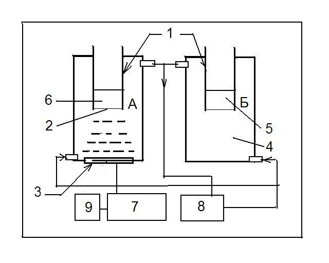
Fig. 1. Installation diagram for exposure to modulated ultrasound: A – thermostatically controlled chamber for ultrasonic exposure, B – thermostatically controlled chamber for control samples. 1 – an internal sample chamber, 2 – a sound–transparent window, 3 – an ultrasonic emitter, 4 – a thermostatic chamber volume, 5 – a control sample, 6 – an irradiated sample, 7 – an ultrasound generator, 8 – an ultrathermostat, 9 - a modulating signal generator. Creatine kinase catalyzes the reaction: creatine + ATF4- --------> creatine phosphat2- + ADP3- + H+ The enzyme was determined in rabbit blood using a diagnostic kit for determining the activity of CORMAY creatine kinase using the kinetic method recommended by DGKC, SFBC and SCE. The method is based on the formation of NADPH initiated by creatine kinase. Creatine Phosphate + ADP <--cc--> creatine + ATP ATP +D-glucose <--hexokinase--> ADP + D-glucose-6-phosphate D-glucose-6-phosphate + NADP + <--G-6-FDG--> D-gluconate-6-phosphate + NADPH + H+ The rate of NADPH formation is directly proportional to the activity of creatine kinase (KK) in the sample. In serum, creatine kinase is stable for up to 10 days at a temperature of 2-8 degrees Celsius or 24 hours at room temperature. The activity was measured spectrophotometrically at a wavelength of 340 nm, in a cuvette with an optical path length of 1 cm. The reaction temperature is 30 ° C, the reaction time is 5 minutes. Absorption values were recorded after 1, 2, 3 minutes. Alkaline phosphatase catalyzes the hydrolysis reaction of p-nitrophenyl phosphate to form colored p-nitrophenol. Phosphatases are specific to the phosphate part of the molecule and can transfer phosphate groups to other molecules, so that the rupture occurs directly behind the phosphorus atom. The clinical significance of alkaline phosphatase is an increase in activity in a number of pathological processes, in particular, in inflammatory processes. A sharp decrease in the activity of alkaline phosphatase is observed in a number of other diseases (radiation sickness, hepatitis, etc.) The activity of alkaline phosphatase in blood serum was determined using a Diakom test kit using a spectrophotometric method. Kinetic spectrophotometric determination of the activity of alkaline phosphatase (alkaline phosphatase) is based on the registration of an increase in the optical density of the prototype at a wavelength of 405 nm as a result of the reaction: DEA, Mg2+ p-Nitrophenyl Phosphate + H2O --------> p-nitrophenol + phosphate Alkaline phosphatase, pH 9.8
The rate of formation of colored p-nitrophenol as a result of the reaction is proportional to the activity of alkaline phosphatase. Blood from Wistar rats was used in the experiments. 2 ml of the reagent solution and 30 µl of the analyzed serum were introduced into the cuvette of the spectrophotometer, stirred and incubated at 37oC for 15 minutes, then 200 µl of the reaction initiator was added, stirred and absorption was measured after 1 min. The measurements were repeated 3 times with an interval of 1 min. The calculation of the change in optical density E per minute was carried out along a rectilinear section of the kinetic curve: For convenience of comparing the results, graphs were plotted in coordinates where relative changes in activity (compared with the control) were postponed on the Y scale, which made it possible to identify active exposure frequencies. The determination of lactate dehydrogenase (LDH) activity is based on a decrease in the optical density of the prototype at 340 nm as a result of the reaction: LDG Pyruvate + NADH + H+ --------> lactate + NAD+ The oxidation of NADH in the presence of LDH is accompanied by a decrease in optical density. To determine the activity of LDH, a set of reagents was used to determine the activity of LDH in serum and plasma "Diakom LDH". Determination of aspartate aminotransferase activity To determine the activity of aspartate aminotransferase (AsAT) standard kits for in vitro diagnostics (AST UV FS) were used. The determination of the activity of AsAT is based on the registration of a decrease in extinction at a wavelength of 340 nm (spectrophotometrically) during the oxidation of NADH. This decrease is proportional to the activity of AsAT in the sample. The standard set of reagents consisted of reagent R1 (tris, pH 7.8, L-aspartate, malate dehydrogenase, lactate dehydrogease), and reagent R2 (NADH, 2-oxoglutarate). The work was performed on rat blood serum taken from the caudal vein. Heparin was used as an anticoagulant when taking blood samples. To determine the control activity of AsAT, 1000 µl of reagent R1 and 200 µl of blood serum were introduced into the cuvette of the spectrophotometer. After 5 minutes of incubation at a temperature of 37 ° C, 250 µl of reagent R2 was added to the cuvette. Extinction was measured after 1 min. for 3 min. In parallel, a sample was placed in the field of modulated ultrasound for a specified time of 5 minutes, after which the extinction value was determined after 1, 2 and 3 minutes. The average value of AsAT activity in the experimental sample was compared with the average value of activity in the control sample. To visually determine the active frequencies, the calculation of the relative change in enzyme activity was carried out according to the formula A =(dE o/dE k -1) * 100%, where dE o and dE k are the changes in optical density in the experiment and control, respectively. Therefore, negative activity values correspond to the amount of suppression of enzyme activity, and positive values correspond to the amount of activation. To determine the activity of blood cholinesterases (CE), standard kits for in vitro diagnostics (from Lachema) were used. The determination of HE activity is based on the registration of an increase in extinction at a wavelength of 405 nm (spectrophotometrically). This increase is proportional to the activity of HE in the sample. The standard set of reagents consisted of reagent R1 (buffer – chromogen, consisting of a phosphate buffer with a pH of 7.9, dithio-bis-nitrobenzoic acid – 40 microns/vial), reagent R2 (substrate – butyrylthiocholiniodide – 1.08 microns/vial) and standard –cholinesterase. The average value of HE activity in the experimental sample was compared with the average value of activity in the control sample. The calculation of the activity change was carried out in the same way as for AsAT. Equipment for exposure to amplitude-modulated ultrasonic waves For ultrasound exposure, a therapeutic ultrasound generator UZT-102 with an external modulation input was used, operating at a frequency of 0.88 MHz in the range of spatial and time average intensities of 0.02 - 1.5 W/cm2 (I SATA – spatial average temporal average intensity – international standard), modulating signal generators (G6-28 and G3-110), a thermostatically controlled chamber for irradiated samples, a digital spectrophotometer for recording the kinetics of an enzymatic reaction (722 Grating Spectrophotometer). When using modulated waves (pulse modulation with a frequency of 2) I SATA – mod = ½ I SATA – not Ultrasound exposure was performed at the beginning of the far zone of the ultrasonic emitter, in the center of the beam, with a diameter of 10 mm in the central spot in the ultrasonic exposure zone. The work uses pulse modulation with a duty cycle equal to 2. This mode allows you to work with equal-energy effects, regardless of the modulation frequency. Metrology of ultrasonic fields
The control of the distribution of ultrasound intensity in the section of the ultrasonic beam in the area of ultrasound exposure to the object under study was carried out: using a differential thermocouple calibrated by ultrasound intensity and the paint/paper method developed by the author of the article using a specially developed program that allows us to represent the intensity distribution in the section of the ultrasonic beam in the form of a three-dimensional image, where the X and Y axes – cross-section dimensions, Z is the value of local intensity or local variable pressure. Thus, it is possible to choose the zone of influence of the ultrasonic field on the object of study with the most uniform amplitude distribution of ultrasound intensities. The program allows you to determine the value of the local intensity at any point in the section of the ultrasonic beam and display the determined points in the table. The nature of the intensity distribution in the same sections of the ultrasonic beam of the same emitter does not change depending on the intensity of ultrasound. Only the degree of staining changes, which is proportional to the intensity in the therapeutic range. The method has a high degree of spatial resolution and image acquisition speed compared to standard methods for monitoring the parameters of ultrasonic emitters and is comparable to optical Schlieren systems. Hydrophones and differential thermocouples average the spatial resolution of intensity distributions over the volume of the sensor used. In our studies, it was important to determine the zone of the ultrasonic beam, the most averaged in intensity, for a more uniform effect on the sample. Modulation does not change the spatial distribution of intensities in a similar section of the field of the emitter used. Only the degree of staining changes, in accordance with the decrease in the spatial and temporal average intensity of modulated ultrasound. Figure 2 shows an example of representing the spatial distribution of ultrasound intensities and determining the values of local ultrasound intensities. 
Fig. 2. An example of a three-dimensional representation of the distribution of local ultrasound intensities in a given section of an ultrasonic beam. Results The results are presented on the graphs in the form of columns with different signs (+/-), since the calculation of the change in activity was carried out using the formula E = (A o / A k - 1) * 100%. Thus, a decrease in activity leads to values with a minus sign in the figure, an increase in activity leads to a plus sign. Such an approach to the analysis of the results allows us to obtain frequency dependences of changes in enzyme activity with a small spread of the data obtained, which do not depend much on the absolute catalytic activity of enzymes changing over time from the moment the object of study is isolated to the end of the experiments. Therefore, when obtaining data, alternating studies according to the control-experiment scheme were used, up to 7 repetitions per one point of the received data for each studied modulation frequency. Figure 3 shows the results of studies of the effect of amplitude-modulated ultrasound with an intensity of I SATA = 0.7 W/cm2 on rabbit blood creatine kinase. It can be seen that the catalytic activity of creatine kinase, regardless of the series, increased when exposed to frequencies of 1-3 Hz and 7-9 Hz and reached a value of 15-80% for these exposure frequencies. A decrease in catalytic activity was observed in the frequency range 3-6 and 10 Hz and reached values from 20-30 % . 
Fig. 3. The dependence of the relative change in creatine kinase activity ((A O – A K) / A K * 100%) on the frequency of ultrasound modulation. Voicing during the reaction for 5 minutes. Differences in the frequency range 1-6, 7-9, 9-10 Hz are significant (P< 0,05). The carrier frequency is 0.88 MHz, I SATA intensity = 0.7 W/cm2. 
Fig. 4. The dependence of the relative change in creatine kinase activity ((A O – A K) / A K * 100%) on the frequency of ultrasound modulation 12 hours after isolation. Differences in the frequency range of 1-4, 5, 6-10 Hz are significant (P<0,05). The carrier frequency is 0.88 MHz, I SATA intensity = 0.7 W/cm2. The frequency dependence of changes in enzymatic activity is observed on freshly isolated preparations. When exposed to long-term storage drugs, the frequency dependence becomes poorly expressed (changes reach only 15-17%), despite maintaining the level of catalytic activity equal to the level of the freshly isolated drug (Fig. 4). Unlike creatine kinase, alkaline phosphatase has a frequency dependence of activity changes when exposed to higher frequencies. Thus, the maximum increase in activity was observed at frequencies close to 350-370 Hz and reached 25%. (Fig. 5), that is, the severity of frequency-dependent changes in enzymatic activity was less than for creatine kinase.

Fig. 5. Dependence of the relative change in the activity of alkaline phosphatase ((A O – A K) / A K * 100%) on the frequency of ultrasound modulation. Voicing during the reaction for 5 minutes (5 measurements for each point). Differences in the frequency range of 300-330, 340-370 Hz are significant (P< 0,05).  A A  B B
Fig. 6. Dependence of the relative change in lactate dehydrogenase activity ((A O – A K) / A K * 100%) depends on the frequency of ultrasound modulation. Voicing of rat blood plasma with pulse-modulated ultrasound for 3 minutes. before the reaction is initiated - A (0.7 W/cm2, 0.88 MHz) and B (0.2 W/cm2, 0.88 MHz) In the frequency range (300 – 400 Hz) The frequency-dependent effects for lactate dehydrogenase differ significantly from those obtained for alkaline phosphatase, both in terms of the magnitude of the effects and in localization of the frequencies of enzyme activation or suppression of its activity (Fig.6A, 6B). We can talk about the multidirectional action of similar frequencies in the same modulation frequency range for two enzymes: alkaline phosphatase and lactate dehydrogenase. It is also seen that when the intensity of ultrasonic exposure changes at the biologically active modulation frequency, the active part of the spectrum of action is transformed and the sign of the effect is changed (Fig.7).  Fig. 7. Dependence of the relative change in lactate dehydrogenase activity on the intensity of modulated ultrasound at a modulation frequency of 400 Hz.  A A  B B Fig. 8. Action spectra for AsAT obtained using AM ultrasound. I SATA= 0.05 W/cm2 (A), I SATA= 0.7 W/cm2 (B), carrier frequency – 0.88 MHz, t volume = 5 min. For aspartate aminotransferase using AM ultrasound for two intensities (0.05 and 0.7 W/cm2) Action spectra were obtained in the modulation frequency range 0-250 Hz (Fig. 8). At an intensity of 0.7 W/cm2, AcAT activation frequencies (70 Hz, 130 Hz, 250 Hz – the maximum activation value) and activity suppression frequencies (110 Hz) were shown. However, with a decrease in the amplitude of the effect, the spectrum of action was transformed and frequencies suppressing the activity of AsAT were revealed A feature of this spectrum of action is the presence of enzyme activity suppression frequencies (modulation frequency – in the range of 88 Hz and 130 Hz), reaching 20-50% activity suppression. And the activation frequency of 130 Hz at ultrasound intensity I SATA = 0.05 W/cm2 is transformed into the frequency of activity suppression at I SATA = 0.7 W/cm2. That is, as in the case of LDH, there is a transformation of the effect sign. For cholinesterase using modulated ultrasound at an intensity of 0.7 W/cm2, an action spectrum was obtained in the modulation frequency range of 0-100 Hz (Fig. 9). 
Fig. 9. Change in the activity of HE under the influence of modulated ultrasound. I=0.7 W/cm2, t = 3 min. Differences in the frequency range of 0-100 Hz are significant (P< 0,05). For HE, frequency-dependent changes in activity were most pronounced compared to other blood enzymes studied. This is the most detailed spectrum of action, taken in 10 Hz increments. The maximum activity changes reached 150-220% at modulation frequencies of 10, 30, 70 and 90 Hz. Discussion of the research results
Frequency-dependent effects of activation and suppression of activity in various frequency ranges of modulation under the influence of modulated ultrasonic waves are observed for almost all studied blood enzymes. This allows us to talk about the universal nature of such effects. As can be seen from Fig. 6 - 8, when modulated ultrasound of the therapeutic intensity range is exposed to blood enzymes, 3 types of effects are observed: 1) when exposed to a modulation frequency that causes at low ultrasound intensities (0.05 W/cm 2) the effect of suppressing activity with an increase in ultrasound intensity, a decrease in the suppression effect is observed, up to zero, and then activation of enzymatic activity and its growth with an increase in ultrasound intensity; 2) when exposed to a modulation frequency that causes at low ultrasound intensities (0.05 W /cm 2) the effect of enzyme activation with increasing intensity, a decrease in the activation effect is observed, up to zero, and then suppression of enzymatic activity and an increase in the suppression effect with increasing ultrasound intensity; 3) the appearance of the activation / suppression effect on some initially neutral frequencies at both low and high intensities of modulated ultrasound. In the first two cases, the transition point of the effect sign is in the area of ultrasound intensity equal to 0.4 W/cm2. The question arises, requiring further research, how such a transformation of the spectrum occurs when the energy of exposure to modulated ultrasound changes, as a result of which activation frequencies become activity suppression frequencies and vice versa. The nonequilibrium dynamic nature of living systems, of which enzyme systems are a part, may be the reason for the dependence of the obtained biological effects of modulated ultrasound exposure on the temporal, frequency and energy parameters of ultrasound. In the study of biological effects, the most significant parameters are usually considered to be the intensity of ultrasound and the total exposure time. Most researchers see the only difference between pulsed and continuous ultrasound only in the change in the ratio between thermal and cavitation effects. Phenomena that may be caused by the dynamics of the biological system and the associated specific effects of modulated influences are usually not taken into account. The effect of ultrasound depends on a number of factors that can be divided into two categories: a) factors related to the influencing agent - ultrasound and b) factors related to the properties of the affected objects. The first category includes such factors of ultrasonic exposure as frequency, intensity, exposure time, energy distribution in the ultrasonic beam, type of modulation, modulation frequencies. The second category for enzyme systems includes such as: acoustic properties of the object, concentrations of enzymes, substrates, in what conformational state are the enzyme molecules, the availability of active enzyme centers for substrate molecules, in what form are the enzyme molecules (tetramers, dimers, monomers), the presence of metal ions in solution, pH of the solution. The formation of ultrasound bioeffects will be determined by a combination of factors of the first and second categories. One of the most important characteristics of the acting ultrasound is the intensity, which determines the nature of the primary physical processes (mechanical, thermal, cavitation) in biological objects located in the ultrasonic beam. Mechanical processes are associated with ultrasound factors such as oscillatory displacement, velocity, acceleration, variable pressure, and their gradients. Thermal processes are associated with the absorption of ultrasonic energy in an object due to relaxation processes in biopolymers. Cavitation processes in the ultrasonic field are caused by the appearance of pulsating gas cavities in the medium, hydrodynamic processes around pulsating cavities and secondary physico-chemical phenomena: mechanical, thermal, electrochemical. The predominance of any of these processes will determine the primary physical mechanism of the biological action of ultrasound. In therapy, depending on the object, ultrasonic waves are used in the range of spatial and temporal average intensities (I SATA) 0.05 - 1.0 W/cm2 (in the USA - 0.05 - 3.0 W/cm2). Due to the spatial heterogeneity of ultrasonic fields, local intensities for flat radiators can exceed the average by several times. For therapy, this is an essential fact, and when choosing the parameters of ultrasonic exposure to a specific object and analyzing the observed effects, it is necessary to take into account the ratio of peak intensity to average. For low-intensity ultrasound (< 0.2 - 0.4 W/cm 2), when thermal and cavitation effects can usually be ignored, the impact on biological objects will be due to mechanical factors. An increase in intensity leads to an increase in the proportion of thermal effects and a qualitative change in the mechanisms of biological action and related effects, for example, temperature changes in enzymatic activity, thermal denaturation of proteins. At even higher intensities, when cavitation phenomena occur, other types of effects are observed, for example, the formation of chemically active compounds that can interact with biomacromolecules. Analysis of the known bioeffects of continuous ultrasound shows that activation processes in biological systems are usually manifested in the zone of mechanical and thermal effects, and inhibition processes begin at the beginning of the cavitation intensity zone. When studying enzymatic reactions and the effects of both continuous and modulated ultrasound on them, a number of mechanisms for increasing or decreasing enzyme activity are observed. It is known that enzymatic reactions occur through the formation of an intermediate product – an "enzyme-substrate complex". In the simplest case:
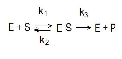
E is an enzyme; k 1 is the rate constant of the reaction of the formation of the enzyme–subrate complex ES. The enzyme-substrate complex can then dissociate with the rate constant - k2 . In addition, the enzyme-substrate complex can be converted into a reaction product P with the release of the enzyme E. The rate constant of formation of the reaction product is P - k 3. The rate constant of a chemical reaction:  Thus, the reaction rate is proportional to the concentration of the ES complex. The increase in the amount of the product depends both on the form in which the enzyme is located in the tetramer-dimer-monomer series and on the conformational state of the enzyme, the more active centers are open to the interaction of the enzyme with the substrate, the higher the yield of the product and vice versa. In addition, the higher the rate of substrate flow to the active center, the higher the rate of formation of the reaction product, the amount of which is used to evaluate the efficiency of the enzyme. Based on this, it can be seen that the effect of modulated ultrasonic waves occurs at the level of processes associated with velocity constants. It should be noted that existing therapeutic ultrasound devices do not allow flexible selection of the frequency of repetition of ultrasound pulses in order to find optimal modes of exposure to a specific organ with a specific pathology. The modes available in therapeutic devices are limited by pulse modulation with a frequency of 50 Hz and pulse durations of 2, 4, 10 msec. It is generally believed that the use of pulse mode in therapy provides a more “mild” effect on tissues, due to a decrease in the total energy injected into the body, and a weakening of thermal effects. However, the main advantage of pulsed ultrasound may be in another way: in the possibility of a more targeted effect on this pathology. Pulse-modulated ultrasound with a pulse repetition rate specially selected for the treatment of a specific disease is not yet used in therapy. It should also be noted that the use of modulated ultrasonic waves in physiotherapy can significantly reduce the energy load on the body and significantly reduce the intensity of the affected ultrasound while maintaining the effectiveness of physiotherapy. Optimization of ultrasound therapy using flexible control of modulation modes is an urgent task of medical acoustics and requires further development. Conclusion The greatest interest in the study of the mechanisms of biological action of physical factors of different nature is caused by the issues of energy and informational action of factors. Energy exposure causes biological effects that qualitatively and quantitatively depend on the amplitude or incident energy of the physical factor. The information component should be understood as the possibility of obtaining various biological effects depending on the modulation frequency with equal-energy and equal-amplitude effects of modulated ultrasound on the biological systems under study, in this case, enzyme systems. Therefore, at the initial stages of research, it is possible to find out in principle the presence or absence of frequency-dependent effects for a given biological system with a fairly rough step. In the case of biological effects, a more subtle dependence of the effects and their correlation with the amplitude-frequency characteristics of the impact factor in the active frequency zone should be clarified, followed by an analysis of functional changes at different levels of the organization of biosystems. Special attention in these experiments should be paid to the same conditions of biosystems and exposure conditions, since the introduction of additional exposure factors, for example, changes in the composition of nutrient media in the study of in vitro systems, additional stress effects that change the functional states of systems in vivo can significantly distort the effects of the active physical factor. n The main approach to the study of modulated waves is to obtain and use action spectra for various biological systems.
n Action spectra – dependences of the magnitude and sign of the biological effect on the modulation frequency with equal-energy and equal-amplitude effects on the system under study. This allows you to control the functional state of biosystems and directionally change the magnitude and sign of the biological effect, since frequencies of multidirectional action are always present in the spectrum. That is, you can use one frequency to get an effect, and use another frequency to either enhance it or completely cancel it. n Using exposure at the active frequency, it is possible to obtain multidirectional effects by changing the energy of the acting ultrasound. Thus, both the modulation frequency and the impact energy can be factors in controlling the functional state of enzyme systems using modulated ultrasonic waves. The main differences between the biological effects of continuous and modulated ultrasound. l The effects of continuous ultrasound depend mainly on the mechanisms of biological action (mechanical, thermal, cavitation). With an increase in the intensity of exposure, one of these mechanisms mainly works. As a rule, with increasing intensity, there is a suppression of the functional state of biosystems at various levels of organization. l The effects of modulated ultrasound may differ significantly from the effects of continuous ultrasound. The presence of active frequencies leads to the transformation of biological effects. At frequencies of activation or suppression of functional activity, effects exceeding the effects of continuous ultrasound can be obtained, with the possibility of changing the sign of the effect with a change in intensity. Thus, it is possible to significantly reduce the intensity of ultrasound exposure in physiotherapy while maintaining the magnitude of biological effects when using modulated ultrasonic fields. It follows from the last point that the thresholds for the development of a biological effect for modulated and continuous ultrasonic waves can be significantly different, that is, the values of the threshold intensities for obtaining a biological effect with continuous ultrasound can be much higher than the threshold intensities for obtaining similar biological effects with modulated ultrasound (I SATA-NON-I SATA-MODES). Thus, the task of the article is to show the presence of frequency-dependent responses of various enzyme systems when exposed to modulated ultrasonic waves of therapeutic intensity range – performed on the example of five enzyme systems. The action spectra for these systems and the possibility of transformation of these spectra with a change in ultrasound intensity are given. It is shown that it is possible to control the magnitude and sign of the responses of the studied enzyme systems, both the modulation frequency and the energy of ultrasonic exposure.
References
1. Grigoriev, Yu.G. (1996). The role of modulation in the biological action of electromagnetic radiation. Radiation biology. Radioecology, 36(5), 659-670.
2. Gapeev, A.B., Sokolov, P.A., & Chemeris, N.K. (2001). Model analysis of the features of the action of modulated electromagnetic fields at the cellular level for various parameters of modulating signal. Biophysics, 46(4), 661-675.
3. Gapeev, A.B., & Chemeris, N.K. (2000). Model approach to the analysis of the effect of modulated electromagnetic radiation on animal cells. Biophysics, 45(2), 299-312.
4. Gapeev, A.B., Yakushina, V.S., Chemeris, N.K., & Fesenko, E.E. (1997). Modulated EMR of extremely high frequencies of low intensity activates or inhibits the respiratory burst of neutrophils depending on the frequency of modulation. Biophysics, 42(5), 1125-1134.
5. Pashovkina, M.S., & Akoev, I.G. (1996). Effect of pulse-modulated microwave radiation at 2375 MHz on the ATPase activity of rat muscle actomyosin. Radiation Biology. Radioecology, 36(5), 700-705.
6. Pashovkina, M.S., & Akoev, I.G. (2000). Changes in the activity of alkaline phosphatase in the blood serum of guinea pigs in vitro under the action of an amplitude-modulated microwave electromagnetic field of 2375 MHz. Biophysics, 45(1), 130-136.
7. Pashovkina, M.S., & Akoev, I.G. (2001). Influence of microwave EMR intensity on the direction and severity of the reaction of alkaline phosphatase in blood serum under weak amplitude-modulated effects // Radiation Biology. Radioecology, 41(1), 62-66.
8. Pashovkina, M.S., Akoev, I.G. (2000). Study of changes in the activity of aspartate aminotransferase in human blood serum under low amplitude-modulated microwave EMR exposure. Radiation biology. Radioecology, 40(6), 700-702.
9. Pashovkina, M.S., Akoev, I.G., & Pashovkin, T.N. (2002). Changes in the activity of some animal and human enzymes under the influence of modulated microwaves and the phenomena of revealed nonlinear effects. In the book. Biological effects of weak electromagnetic radiation. Pushchino, 26-37.
10. Gapeev, A.B., & Chemeris, N.K. (1999). Effect of continuous and modulated EMR EHF on animal cells. Review. Part I. Features and main hypotheses about the mechanisms of biological action of EMR EHF. Bulletin of new medical technologies, 1, 15-22.
11. Svidovy, V.I., Kolmakov, V.N., & Kuznetsov, G.V. (1985). Changes in the activity of aminotransferases and the permeability of erythrocyte membranes under the influence of infrasound and low-frequency noise. Hygiene and Sanitation, 10, 73-74.
12. Svidovy, V.I., Kolmakov, V.N., Kuleba, V.A., & Timofeeva, V.M. (1987). Changes in permeability, total ATP-ase activity of erythrocytes and superoxide dismutase activity of blood plasma under the influence of infrasound. Hygiene and sanitation, 5, 78-79.
13. Alekseev, S.V., Kolmakov, V.N., & Svidovy, V.I. (1984). Influence of low-frequency acoustic vibrations on some properties of erythrocyte membranes. Hygiene and sanitation, 2, 82-84.
14. Kolmakov, V.N., Svidovy, V.I., & Shleikin, A.G. (1984). Influence of low-frequency acoustic oscillations on some components of erythrocyte membranes. Occupational Health and Occupational diseases, 10, 48-49.
15. Novikov, A.M. (1976) Histochemical studies of enzymatic-metabolic changes in striated muscle fibers under the action of infrasound. Proceedings of LSGMI, 114, 30-32.
16. Gabovich, R.D., Shutenko, O.I., & Krechkovsky, E.A. (1984). Influence of infrasound on bioenergy processes, ultrastructural organization of organs and some regulatory processes. Hygiene and Sanitation, 3, 9-15.
17. Barseghyan, V.O. (1981) Investigation of the effect of chemical agents and ultrasound on the activity of alkaline phosphatase. PhD dissertation, Yerevan.
18. Svidovy, V.I., & Shleikin, A.G. (1987). On the effect of infrasound on the activity of tissue succinate dehydrogenase. Occupational health and occupational diseases, 5, 50-52.
19. Goncharova, L.P., Kadyskina, E.N., Makarova, I.N., & Rodionova, L.P. (1984). Comparative study of the effect of noise of different intensity on some components of the antioxidant system of the cell. Antioxidants and Adaptation, L., 22-26.
20. Melkoyan, M.M., & Melik-Agaeva, E.A. (1984). Influence of noise on the processes of lipid peroxidation. Biol. Journal of Armenia, 37(8), 668-677.
21. Alekseev, S.V., Svidovy, V.I., & Velichko, L.N. (1983). Influence of low-frequency acoustic vibrations on the phospholipid composition of whole blood and some animal tissues. Occupational health and occupational diseases, 3, 39-41.
22. Sarvazyan, A.P., Belousov, L.V., Petropavlovskaya, M.N., & Ostroumova, T.V. (2002). The action of low-intensity pulsed ultrasound on amphibian embryonic tissues. Ultrasound in Med. & biol., 8, 639-654.
23. Melnikova, E.V., Uteshev, V.K., Pashovkin, T.N., Sokolova, A.A., Yashin, V.A., Karnaukhov, V.N., & Gakhova, E.N. (2006). Change in the permeability of amphibian embryonic membranes for fluorescent dyes under the action of ultrasound. Biophysics, 3, 500-504.
24. Pashovkin, T.N. (2005). Biological effects of amplitude-modulated ultrasonic fields in the therapeutic range of intensities. On Sat. materials of the II Eurasian Congress on Medical Physics and Engineering "Medical Physics-2005", Moscow, June 21-24, 2005, 223.
25. Pashovkin, T.N., & Pashovkina, M.S. (2005). Effects of amplitude-modulated therapeutic ultrasound on blood enzymes. 5TH International Symposium on Therapeutic Ultrasound. abstracts. October 27–29 October, 2005, Harvard Medical School, Boston, T063, P. 39.
26. Gakhova, E.N., Pashovkin, T.N., Melnikova, E.V., Uteshev, V.K., & Sadikova, D.G. (2007). Ultrasound can alter the permeability of amphibian embryonic membranes. Veterinary pathology, 1, 7-9.
27. Uteshev, V.K., Pashovkin, T.N., & Gakhova, E.N. (2010). Survival of amphibian embryos after exposure to modulated ultrasound in the therapeutic range of intensities. Bulletin of new medical technologies, 4, 7-10.
28. Oleshkevich, A.A., & Pashovkin, T.N. (2014). Quantitative analysis of the effect of modulated ultrasound on some animal tissue cells. Veterinary medicine, zootechnics and biotechnology. Scientific and practical journal. FGBOU VPO MGAVMiB, 5, 27-33.
29. Oleshkevich, A.A., & Pashovkin, T.N. (2015). Effect of modulated ultrasound in Natrinema Pallidum (Halobacterium halobium) and Avilibrio fisheri. Mater. International Symposium "MICROORGANISMS AND BIOSPHERE" MICROBIOS-2015, November 25-30, 2015 Tashkent, Uzbekistan.
30. Oleshkevich, A.A., Vasilevich, F.I., & Pashovkin, T.N. (2015). Biochemical and biophysical effects of continuous and modulated ultrasonic waves on Alivibrio fisheri and Natrinema pallidum. Veterinary, zootechnics and biotechnology, 12, 50-56.
31. Gapeyev, A.B., Lysenko, Yu.N., & Pashovkin, T.N. (2016). Combined drug and ultrasound on cancer treatment. // Invited oral presentation at the International Conference on "New Approaches fighting Cancer and Aging", December 2-3, 2016 in Nanjing, China.
32. Pashovkin, T.N., Shilnikov, G.V., & Sarvazyan, A.P. (1986). Ultrasonic field visualization method. A.s. No. 1206693 . B.i. No. 3, 01/23/86.
33. Pashovkin, T.N., & Shilnikov, G.V. (2000) Registration and analysis of intensity distributions in ultrasonic beams using dyes. Scientific Instrumentation, 10(3) 17-26.
34. Sadikova, D.G., & Pashovkin. T.N. (2005). Intensity distribution in cross sections of ultrasonic beams of therapeutic and diagnostic emitters in liquid media. On Sat. materials of the II Eurasian Congress on Medical Physics and Engineering "Medical Physics-2005", Moscow, June 21-24, 2005, p. 225.
35. Pashovkin, T.N., Pashovkina, M.S., Sadikova, D.G., & Shilnikov, G.V. (2006). Intensity distribution in ultrasonic beams of therapeutic transducers using dyes: 3D representation of intensity distributions in ultrasonic beam cross sections and 3D reconstruction of ultrasonic fields in aqueous media. Bulletin of new medical technologies, 3, 155-159.
36. Bawin, S.M., Gavalas-Medici, R., & Adey, W.R. (1973). Effects of modulated very high frequency fields on specific brain rhythms in cats. Brain Res, 58, 365-384.
37. Bawin, S.M., Kaczmarek, L.K., Adey, W.R. (1975). Effects of modulated VHF fields on the central nervous system. Ann NY Acad Sci, 247, 74-80.
38. Blackman CF, Elder JA, Weil CM, et al. (1979). Induction of calcium ion efflux from brain tissue by radio frequency radiation: effects of modulation frequency and field strength. Radio Sci, 14, 93-98.
39. Blackman, C.F., Benane, S.G., House, D.E., et al. (1985). Effects of ELF (1–120 Hz) and modulated (50 Hz) RF fields on the efflux of calcium ions from brain tissue in vitro. Bioelectromagnetics, 6, 327-338.
40. Byus, C.V., Lundak, R.L., Fletcher, R.M. et al. (1984). Alterations in protein kinase activity following exposure of cultured lymphocytes to modulated microwave fields. Bioelectromagnetics, 15, 217-238.
41. Litovitz, T., Krause, D., Penafiel, M., et al. (1993). The role of coherence time in the effects of microwaves on ornithine decarboxylase activity. Bioelectromagnetics, 14, 395–404.
42. Dalecki, D., Raeman, C.H., Child, S.Z. & Carstensen, E.L., (1995). Intestinal hemorrhage from exposure to pulsed ultrasound. Ultrasound in medicine & biology, 21(8), 1067-1072.
43. [Bialek and Wit, 1984] Bialek W, Wit HP (1984): Quantum limits to oscillator stability: theory and experiments on acoustic emissions from the human ear. Phys Lett N 104A, 173-178.
44. Lennart, D. Johns (2002). Nonthermal Effects of Therapeutic Ultrasound: The Frequency Resonance Hypothesis. Journal of Athletic Training, 37(3), 293-299.
45. Pinamonti, S., Gallenga, P.E., & Masseo, V. (1982). Effects of pulsed ultrasound onhuman erythrocytes in vitro. Ultrasound Med Biol., 8, 631-638.
46. Nykanen, M. (1995). Pulsed ultrasound treatment of the painful shoulder: a randomized, double-blind, placebo-controlled study. Scand J Rehabil Med, 27, 105-108.
47. Dyson, M. (1987). Mechanisms involved in therapeutic ultrasound. Physiotherapy, 73, 116-120.
48. Kitchen, S.S., & Partridge, C.J. (1990). A review of therapeutic ultrasound. Physiotherapy, 76, 593-600.
49. Nilsson, A.M., Odselius, R., Roijer, A., Olsson, S.B. (1995). Pro- and antifibrinolytic effects of ultrasound on streptokinase-induced thrombolysis. Ultrasound Med Biol., 21, 833-840.
50. Ter Haar, G. (1999). Therapeutic ultrasound. Eur J Ultrasound, 9, 3-9.
Peer Review
Peer reviewers' evaluations remain confidential and are not disclosed to the public. Only external reviews, authorized for publication by the article's author(s), are made public. Typically, these final reviews are conducted after the manuscript's revision. Adhering to our double-blind review policy, the reviewer's identity is kept confidential.
The list of publisher reviewers can be found here.
The reviewed article is devoted to frequency-dependent changes in the activity of a number of key blood enzymes in laboratory animals under the influence of modulated ultrasonic waves. For continuous ultrasound used in medicine for physiotherapy and phonophoresis of medicinal substances, a fairly large number of works have been presented in the world literature describing biological effects and mechanisms of biological action, including mechanical, thermal and cavitation mechanisms. This is due to the safe use of ultrasonic waves. Therefore, the main effects are described for a range of spatial and temporal ultrasound intensities of 0.05 – 3.0 W/cm2 and exposure times up to 15 minutes. At the same time, it is noted in a number of works that for modulated ultrasound with various types of modulation, biological effects can be observed that exceed the effects of continuous waves at a lower (at least twice) intensity. That is, the thresholds for the occurrence of biological effects of modulated ultrasound may be significantly lower than those caused by continuous ultrasound. There is also another difference between the biological effects of continuous and modulated waves – the non–specificity of the biological effects of continuous waves and the specificity for modulated waves, expressed in the presence of frequency-dependent effects. In the latter case, the effects may differ at different modulation frequencies, but with equal energy effects on the biological systems under study. That is, in addition to the basic mechanisms operating under the influence of continuous waves, there are mechanisms of action associated with the use of wave modulation. This is why the relevance of research on the mechanisms of biological action of modulated ultrasonic waves is connected. In addition, the use of modulation modes when exposed to ultrasound can increase the efficiency and selectivity of ultrasound application in medicine while reducing the energy of the affected ultrasound. That is, to increase the safety of ultrasound in medicine. This article is structured in a traditional style, contains an introduction with a literature review, materials and methods, results, discussion of results, conclusion and a list of used literature. The introduction provides a review of the literature, which provides data on frequency-dependent effects when modulated waves are applied to biological systems at different levels of organization. The concept of action spectra and the importance of obtaining them under the equal-energy action of modulated waves to identify biologically active frequencies for the studied objects are considered. Special attention is paid to the metrological support of experiments with ultrasound, since in the near and early far zones of the emitters used, the distribution of local intensities in the sections of ultrasonic beams is heterogeneous. This is illustrated in Figure 1. The reproducibility of the results depends on the correct metrology. The paper presents frequency-dependent changes in enzyme activity for five different blood enzymes when exposed to modulated ultrasonic waves of therapeutic intensity range. It is shown that the action spectra can vary depending on the intensity of ultrasound. Thus, it was shown that the controlling factors shaping biological effects can be both modulation frequencies and the energy factor (ultrasound intensity). That is, the use of modulated ultrasonic waves in medicine can be targeted. To date, there is no theory of the effect of modulated ultrasonic waves on biological systems of different levels of organization. Therefore, obtaining correct data on frequency-dependent biological effects is a prerequisite for the development of such a theory (or theories). The presentation of data on the frequency-dependent change in enzyme activity in the form of a relative change in activity allows us to use the data obtained by the author from various experiments with different initial levels of enzyme activity and increase the reproducibility of the results, with the determination of biologically active frequencies. The data presented in the article are beyond doubt, since they are based on a sufficient amount and high quality of scientific material. The list of cited literature contains 35 publications. Summarizing the above, we can say that the article "Frequency-dependent changes in the activity of blood enzymes under the influence of modulated ultrasound" should be published in the scientific journal "Physics of Biology and Medicine".
|

 Eng
Eng


